In a study more than a decade in the making, CSUN biologists have compared the fine-scale neuroanatomy of two nematode worms to describe evolutionary changes in their simple nervous systems since they last shared a common ancestor, over 100 million years ago. The study has been recently published in the journal eLife.
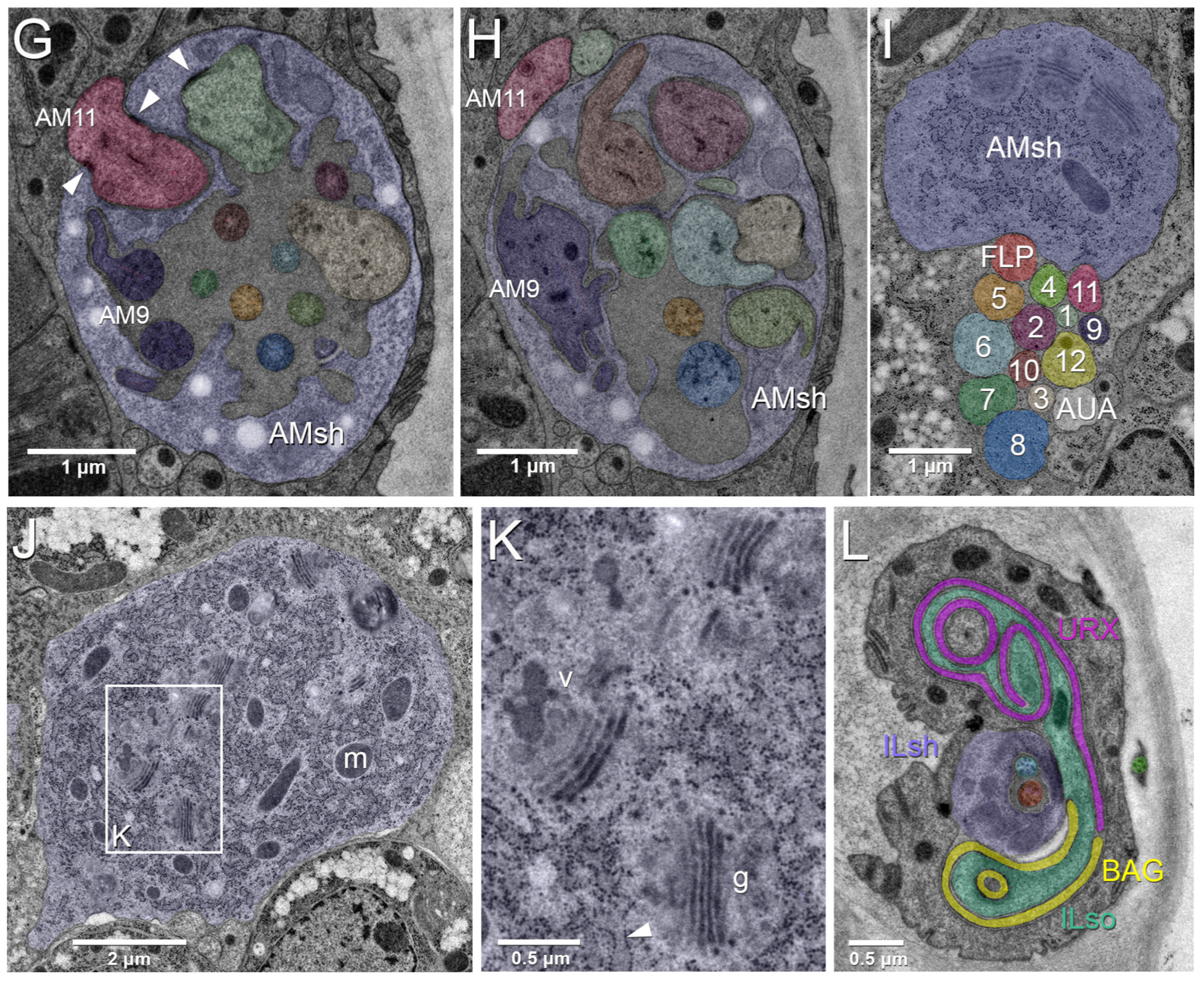
Professor of Biology Ray Hong and undergraduate researchers Heather Carstensen and Jessica Castrejon worked with collaborators in at the Max Planck Institute, Columbia University, and the Howard Hughes Medical Institute to use electron micrographs obtained over a decade ago in reconstructing the ultrastructures of sensory neurons in Pristionchus pacificus, a roundworm that lives symbiotically with scarab beetles. Pristionchus pacificus is in the same order as the widely used laboratory model Caenorhabditis elegans, sharing a common ancestor with the more widely-studied worm more than 100 million years ago — a greater evolutionary divide than that separating humans from mice.
Comparing the P. pacificus micrographs to the fine-scale neuroanatomy of C. elegans revealed and found major changes in connections among sensory nerve cells, but not in the total number of sensory nerve cells. The authors call the similarities between the two worms remarkable, given their differing lifestyles and their long history of independent evolution.
The full paper is available open access from the journal website.
Image: Electron micrograph images of sections through the amphid sensillum of P. pacificus (from Hong et al 2019, Figure 1)

